Unusual Papillary Lymphoid Hyperplasia of The Palatine Tonsil Associated with Ebstein Barr Virus Infection in a Child: A Case Report
Andrea Borromini*, Alessandro Marchesi, Gloria Golinelli, Maurizio Salvadore, Mascia Verzeletti, Francesca Andrea Bonarrigo, Nicolas Martinelli, Michela Todeschini and Gianpaolo Mirri
DOI10.21767/2471-805X.100021
Andrea Borromini1*,Alessandro Marchesi2, Gloria Golinelli2, Maurizio Salvadore4, Mascia Verzeletti5, Francesca Andrea Bonarrigo6, Nicolas Martinelli1, Michela Todeschini7 and Gianpaolo Mirri3
1Department of Internal Medicine,Faculty of Medicine and Surgery, University of Pavia, Italy
2Department of Otorhinolaryngology,Hospital Sant’Antonio Abate, Gallarate,ASST Valle Olona, Italy
3Department of Pediatric Care, Saronno Hospital, ASST Valle Olona, Italy
4Department of Anatmopathology, Hospital Sant’Antonio Abate, Gallarate, ASST Valle Olona, Italy
5Department of Pediatric Care, Hospital Sant’Antonio Abate, Gallarate, ASST Valle Olona, Italy
6Pediatric Highly Intensive Care Unit, Department of Pathophysiology and Transplantation, University of Milan, Fondazione IRCCS Ca 'Granda Ospedale Maggiore, Italy
7Agency for the Health Protection, Milan - Metropolitan City, Italy
- *Corresponding Author:
- Andrea Borromini
Department of Internal Medicine
Faculty of Medicine and Surgery
University of Pavia, Italy
Tel: +393392358517
Fax: +278615107002
E-mail: andrea.borromini01@ universitadipavia.it
Received date: November 23, 2016; Accepted date: December 05,2016; Published date: December 12, 2016
Citation: Borromini A, Marchesi A, Golinelli G, et al. The Ebstein-Barr Virus as the Triggering of Papillary Lymphoid Hyperplasia of the Palatine Tonsil in a Child:A Case Report. J Pediatr Care. 2016, 2:3. doi:10.21767/2471-805X.100021
Abstract
We describe the case of an Italian, Caucasian nine-year-old girl, suffering from a unilateral tonsillar swelling. Histopathological and immune-histochemical exams show a lymphoepithelial reactive hyperplasia overlap with a chronicle inflammation.
Microbiologic swaps reveal a throat infection caused by Streptococcus pyogenes, and a probable viral infection, caused by Ebstein Barr Virus (EBV). We underline the importance to recognise and consider this benign lesion due to its similarities with a malignant neoplastic form, especially during differential diagnosis of cervical swellings in young women. This is one of the rare cases described in scientific literature in the last years, especially among caucasian race.
https://marmaris.tours
https://getmarmaristour.com
https://dailytourmarmaris.com
https://marmaristourguide.com
https://marmaris.live
https://marmaris.world
https://marmaris.yachts
We describe the case of an Italian, Caucasian nine-year-old girl, suffering from a unilateral tonsillar swelling. Histopathological and immune-histochemical exams show a lymphoepithelial reactive hyperplasia overlap with a chronicle inflammation.
Microbiologic swaps reveal a throat infection caused by Streptococcus pyogenes, and a probable viral infection, caused by Ebstein Barr Virus (EBV). We underline the importance to recognise and consider this benign lesion due to its similarities with a malignant neoplastic form, especially during differential diagnosis of cervical swellings in young women. This is one of the rare cases described in scientific literature in the last years, especially among caucasian race.
Keywords
Tonsillar hyperplasia; EBV; HPV; Malignancies; Lymphoid proliferation
Introduction
A nine-year-old Caucasian Italian girl was referred to our Pediatric Clinic due to some episodes of vomiting with blood traces and loss of appetite, occurred in the two days before. She presented in good general condition, pale skin, but normal vital signs; during thoracic and abdominal examination, no pathological findings were noted; her weight was 20.8 Kg. A submandibular lateral adenopathy associated to a multi nodular lesion of the right palatine tonsil, of about 2 cm, were noticed during the cervical physical examination, without any risk for obstruction of the upper airway.
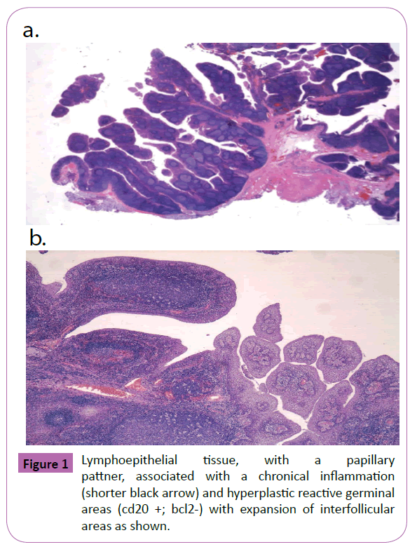
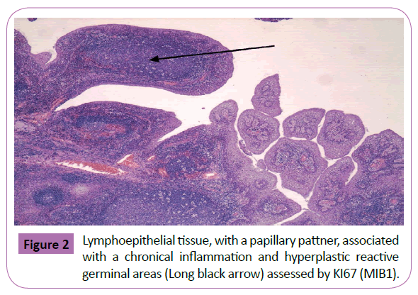
Blood tests showed a moderate leukocytosis without inflammation index (WBC 16, 300/mm3, CRP 0.8 mg/dL, ESR 25 mm/h) and the throat swab indicated a streptococcal infection, sensitive to penicillin and an antibiotic therapy with ampicillinsulbactam was began. After that, a blood serological research for IgG and IgM antibodies against Cytomegalovirus was performed, resulting negative. EBV-VCA and EBV-EBNA IgG antibodies indicated a prior Epstein Barr Virus (EBV) infection with a faint positivity for EBV IgM antibody. No alterations to electrophoresis were founded. No pulmonary alterations were found at the chest X-ray. Due to the possibility of a malignant neoplastic form, a tonsillectomy “secondo Rose” was done. The palatine tonsil macroscopically appeared papillary, brownish with hemorrhagic areas; instead, microscopically it presented a lymphoepithelial tissue, with a papillary pattner, associated with a chronical inflammation and hyperplastic reactive germinal areas (CD20 +; Bcl2-) with expansion of interfollicular areas (Cell. Bcl2 + CD3 + T) (Figures 1 and 2). The proliferation index, assessed with Ki67 (MIB1), was founded in the 20% of the cells outside these germinal areas. The research of 18, 18, 31, 33, 35, 39, 45, 51, 52, 56, 58, 66 genotypes Human Papillomavirus (HPV), performed by in situ hybridisation, was negative. The child was discharged on the tenth day, considering the absence of surgical complications.
Discussion
We wanted to present this case report to the scientific community in order to contribute to spread the knowledge about this unusual clinical condition, rarely described until now: the linfopapillary tonsillar hyperplasia.
It is useful to discuss this rare case of benign hyperplasia for the follow reasons.
1. First of all, this clinical condition is typically described in Japan population [1] and its manifestation is sporadic in Caucasian people [2,3]. We could suppose there is a correlation between the onset of this disease and an external trigger (viral and/or environmental factor), predisposing to the development of a reactive form of aberrant lymph node germinal centers. The Immuno- Mediated Lymphocyte T response may represent the main way for pathological proliferation, but nowadays the exact mechanism is already unclear. Clinically, the majority of unilateral cervical neoformations - as wrote by Zhao et al. [4] is malignant. Anyway, we must consider that the palatine tonsil could physiologically represents the reactive proliferation response to an inflammation process of the upper throat, transforming its micro pattern into a lymphoid one.
2. It is important, during cervical examination, to put the attention on the probability of a repentine obstruction of the upper airways, as showed by Carillo-Faga [2]. The literature documents the possible autosomal dominant transmission of this disease and the correlation between viral infections (EBV and HPV) and the development of nasopharyngeal tumors [5-7], so that, we made the exams to individuate the presence of EBV or HPV.
3. Secondly, the positivity for EBV and Streptococcus pyogenes, highlights the link between external triggers and chronic inflammation response, bringing, in individual with a predisposition, to a proliferative reaction and remodeling in the sense of papillary tissue pattern and polyclonal T Lymphocytes expansion that boosts the aberrant hypertrophy.
4. Finally, we conclude with a remark: Kaneko et al. describe as an atypical tonsillar hyperplasia may resemble a mucous EBV-related lymphoma [7]. Could this rare disease be considered as a pre-cancerous lesion? In this case tonsillectomy and pathological study would represent the gold standard treatment.
Take Home Message
This case indicates the importance of an accurate differential diagnosis in tonsillar hypertrophy, especially if there are evidences of infectious or inflammatory condition.
It is possible that EBV acts as a trigger determining aberrant proliferation for tonsillar reactive hyperplasia. Even if the clinical patient’s history was unremarkable, it is important to perform virologic tests, to exclude EBV or HPV infections. Particularly, the exclusion of HPV infections is essential, for the surgery follow-up and for medical and legal aspects related to pediatric patients. Indeed, we suggest to analyzed the hyperplasia, after surgical therapy, to rule out malignant form, especially in Asiatic patients, without excluding it “ex ante” in Caucasian ones.
References
- EnomotoP (1980)Papillary hypertrophy of the palatine tonsils. Ann OtolRhinolLaryngol 89: 132-134.
- Carrillo-Farga J (1983)Lymphoid papillary hyperplasia of the palatine tonsils. Am J Surg Pathol 7: 579-582.
- Dias G (2003)Lymphoid papillary hyperplasia: Report of a case. Oral Surg Oral Med Oral Pathol Oral RadiolEndod 95: 77-79.
- Zhao Y (2013)Lymphoid papillary hyperplasia of the palatine tonsil: A Chinese case report. Int J ClinExpPathol 6: 1957–1960.
- PathmanathanS (1995)Undifferentiated, non-keratinizing, and squamous cell carcinoma of the nasopharynx. Variants of Epstein-Barr virus-infected neoplasia. Am. J. Pathol.146:1355-1367.
- Mallen-St Clair J (2016) Human papillomavirus in oropharyngeal cancer: The changing face of a disease.Biochim Biophys Acta 1866:141-150.
- Kaneko Y (2014)Atypicalinterfollicular hyperplasia of tonsils resembling mucosa-associated lymphoid tissue lymphoma: A clinic-pathological, immuno-histochemical study and Epstein-Barr virus findings in 12 cases. J Clin Exp Hematop.
Open Access Journals
- Aquaculture & Veterinary Science
- Chemistry & Chemical Sciences
- Clinical Sciences
- Engineering
- General Science
- Genetics & Molecular Biology
- Health Care & Nursing
- Immunology & Microbiology
- Materials Science
- Mathematics & Physics
- Medical Sciences
- Neurology & Psychiatry
- Oncology & Cancer Science
- Pharmaceutical Sciences


