Neurenteric Cysts at Foramen Magnum in Children
Arnold H Menezes*
Arnold H Menezes*
Department of Pediatric Neurosurgery, University of Iowa Hospitals and Clinics, Iowa City, Iowa 52242, USA
- *Corresponding Author:
- Arnold H Menezes
Department of Pediatric Neurosurgery, University of Iowa Hospitals and Clinics, Iowa City, Iowa 52242, USA
Tel: +319-356-2768
E-mail: arnold-menezes@uiowa.edu
Received Date: August 18, 2020; Accepted Date: September 01, 2020;Published Date: September 07, 2020
Citation: Menezes AH (2020) Neurenteric Cysts at Foramen Magnum in Children. J Pediatr Care Vol.6 No.3:5. DOI: 10.36648/2471- 805X.6.3.100050.
Description
In the late 19th century an interesting condition was recognized in autopsies in several aborted fetuses that exhibited a variable syndrome of related developmental defects in the vertebral column, spinal cord and the gastrointestinal tract [1,2]. This can result in cysts, fistulae and spinal dysraphic states. Neurenteric cysts (NEC) can be formed within the chest, abdomen, brain and the spine.
Nervous system neurenteric cysts commonly present in the first two decades of life. Several case reviews have been done [1-4]. The diagnosis of neurenteric cyst is based on the location and the findings of a cystic cavity lined by cuboidal or columnar epithelial with mucin producing droplets. Positive immunoreactivity of tissue for carcinoembryonic antigen suggest an endodermal origin and has been used to assist in the diagnosis. These cysts show presence of keratin markers. The embryogenesis of lesions described as neurenteric cyst has been approached by numerous authors and we ourselves have described this [3]. They have been termed enterogenous, neurenteric cysts, teratoid cysts, enteric cysts and split notochord syndrome due to location elsewhere in the mediastinum and abdomen. The terminology obviously differs because of the various disciplines involved that view the lesions from different perspectives.
Spinal neurenteric cysts form 94% of the central nervous system neurenteric cysts and an intramedullary form in 14% [3]. There is a male predominance (61%) in the pediatric cases reported and the mean age is 6.4 years with a median of 6 years.
The youngest patient reported with nervous system NEC was a newborn infant who died 70 minutes after birth. In the posterior fossa NEC, 20% are in the pediatric population [1]. The youngest infant in our own experience was diagnosed in utero at 7 months gestation with a large temporal and posterior fossa NEC. In 16 consecutive patients with NEC in the central nervous system in children in another series, 3 were in the posterior fossa and the rest in the spinal canal [2]. Five of the 16 patients had associated anomalies such as the Klippel-Feil syndrome, hemivertebra, diastematomyelia and also neurocutaneous abnormalities. Incomplete resection in 3 led to recurrence.
Neurenteric cysts produce symptoms by their location. This may be acute or chronic onset. The acute deterioration is secondary to motion of the cyst against distorted vascular and neural structures and at times due to hemorrhage. Neurenteric cysts may present as recurrent meningitis.
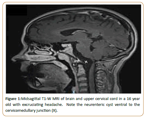
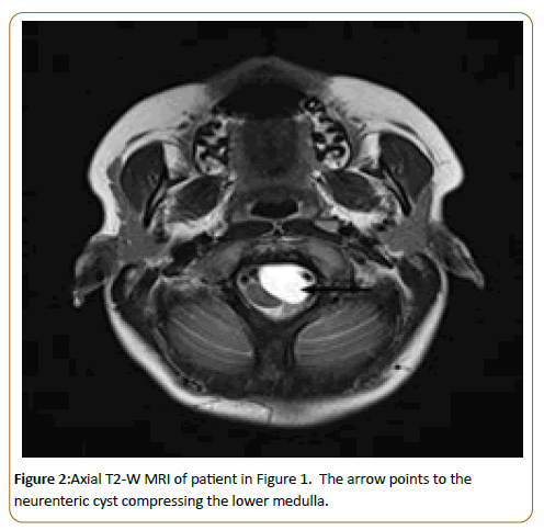
Neurenteric cysts of the posterior fossa have been reviewed by us. Most recently five of these were in children, located ventral to the medulla, straddled by the vertebrobasilar arterial tree [4]. The recent report also reviews that in the literature. Headaches may resemble migraines but at times can be excruciating as in the cases described by us. Intracranial neurenteric cyst should be considered in the differential diagnosis for any well demarcated extra-axial cystic lesion below the tentorium. The cysts are isointense to hyperintense on MRI T1-W imaging and variable T2 signal intensity Figures 1 and 2. They may or may not show enhancement. The risk of rapid expansion, hemorrhage and neurological deficit and the location in a confined space in the posterior fossa calls for surgical excision.
The treatment of neurenteric cyst is complete excision. When present in the spinal canal or at foramen magnum, they are located anterior to the dentate ligaments and closely associated with vascular structures which are distorted and can contribute to neurological symptoms.
Surgical treatment is curative.
References
- Bejjani GK, Wright DC, Schessel D, Sekhar LN (1998) Endodermal cysts of the posterior fossa. Report of three cases and review of the literature. J Neurosurg 89: 326-335.
- De Oliveira RS, Cinalli G, Roujeau T, Sainte-Rose C, Pierre-Kahn A et al. (2005) Neurenteric cysts in children: 16 consecutive cases and review of the literature. J Neurosurg 103: 512-523.
- Menezes AH, Traynelis VC (2006) Spinal neurenteric cysts in the magnetic resonance imaging era. Neurosurgery 58: 97-105.
- Menezes AH, Dlouhy BJ (2020) Neurenteric cysts at foramen magnum in children: Presentation, imaging characteristics and surgical management-Case series and literature review. Childs Nerv Syst 36: 1379-1384.
Open Access Journals
- Aquaculture & Veterinary Science
- Chemistry & Chemical Sciences
- Clinical Sciences
- Engineering
- General Science
- Genetics & Molecular Biology
- Health Care & Nursing
- Immunology & Microbiology
- Materials Science
- Mathematics & Physics
- Medical Sciences
- Neurology & Psychiatry
- Oncology & Cancer Science
- Pharmaceutical Sciences


