A Case of Neonatal Femur Shaft Fracture in Twin Prenancy during Caeserean Section
Chetan John Rasquinha , Ghanaprakash Pallaniappan, Sutharsen NR, Major K Kamalanathan and Hubert Praveen Abner
Chetan John Rasquinha*, Ghanaprakash Pallaniappan, Sutharsen NR, Major K Kamalanathan and Hubert Praveen Abner
Department of Orthopaedics, PSG Institute of Medical Sciences & Research, Coimbatore, Tamil Nadu, India
- *Corresponding Author:
- Chetan John Rasquinha
Department of Orthopedics,
PSG Institute of Medical Sciences and Research,
Coimbatore, Tamil Nadu, India,
Tel: 944902598;
E-mail: chetangemini85@gmail.com
Received Date: November 11, 2020; Accepted Date: November 21, 2020;Published Date: November 28, 2020
Citation: Rasquinha CJ, Pallaniappan G, Sutharsen NR, Kamalanathan MK, Abner HP (2020) A Case of Neonatal Femur Shaft Fracture in Twin Pregnancy during Caeserean Section. Pediatric Care Vol.6 No.4: 2
Abstract
Femoral shaft in twin pregnancy of new-borns by caesarean section is rare and not a common occurrence. Literature has highlighted long bone fractures to be very common in virginal delivery compared to caesarean section. Foetal injuries complicated 1.1%. In a multicentre randomised study by Hannah et al. showed that the incidence of fracture of long bones in caesarean section is 0.1% and 0.5% in vaginal delivery.
Hence caesarean section reduces the incidence of femur fractures, however some difficult extractions and manoeuvres employed during caesarean section can lead to femur fractures in neonates.
Our case report is a twin pregnancy of 1st twin (girl) in transverse lie and the other 2nd twin (boy) in breech with cord around the neck which was taken up for emergency C-section in view of foetal growth retardation grade 3 (FGR) of 2nd twin with reversed end diastolic flow (EDF) in the 1st twin.
Keywords
Neonate; Birth injuries; Femoral shaft fractures; Caesarean transverse lie
Introduction
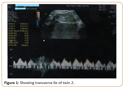
Femur shaft fractures following birth in new-borns is a very rare injury [1-5]. Birth injuries occurring due to trauma and process of child birth in vaginal breech deliveries are most common. The role of caesarean delivery in reducing the outcome of these injuries is a matter of debate [6,7]. We report a case of dizygotic twin pregnancy of new-born delivered by Lower Segment Caesarean Section (LSCS) for transverse lie foetal presentation (Figure 1), producing right femoral shaft fracture in female twin treated with Pavlik harness.
Case Report
A 2 days old infant was referred to the orthopaedic department at PSGIMSR from the neonatal ICU for complaints of swelling, deformity and inability to move right leg from the time of birth. The neonate was crying incessantly.
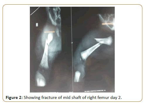
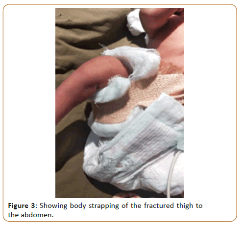
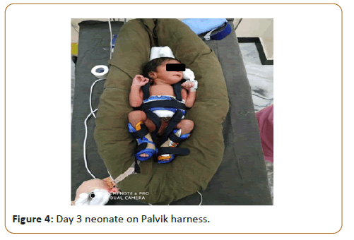
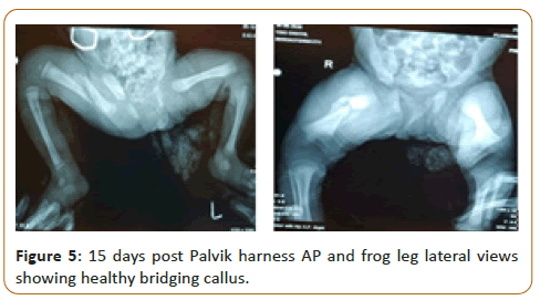
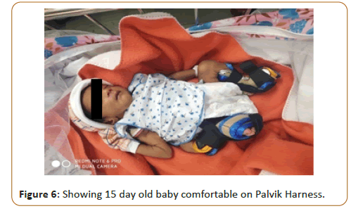
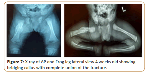
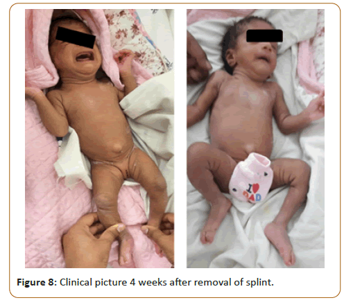
On examination there was swelling and abnormal mobility in the right thigh. X ray revealed a fracture of the mid shaft of the femur with flexion and abduction of the proximal fragment. Initially was managed with body strapping, thigh approximated to the abdomen (Figure 2), so that the distal fragment was aligned with the proximal fragment. Since this strapping was cumbersome, it was interfering with the hygienic care of the abdomen and perineum we followed this up with the application of a Pavlik harness which is usually used to maintain the position of flexion and abduction in CDH infants. This splint by keeping the limb in flexion and abduction helped in the anatomical alignment of the distal fragment to the proximal and it was patient friendly. Palvik harness is a useful adjunct in the management of neonatal femur shaft fractures (Figures 3-8).
Results and Discussion
The earliest case of femoral shaft fracture in a new-born was reported in 1922 following a difficult breech delivery [7,8]. In our case of twin pregnancy being an emergency LSCS due to Foetal Growth Retardation grade 3 (FGR) of one twin with reversed End Diastolic Flow (EDF) in the same twin. The need of considerable traction and the extraction manuveour can lead to such femur fractures.
The potential for bone remodelling and that any angulation at the time of fracture union will remodel over time in neonates. Palvik harness which is usually used to maintain the position of flexion and abduction in CDH infants. This splint by keeping the limb in flexion and abduction helped in the anatomical alignment of the distal fragment to the proximal and it was patient friendly less invasive for neonates. This treatment provides a onetime application with excellent clinical outcomes. Though this is a rare presentation must be looked out for especially in twin pregnancy Caesarean sections. Early presentation as in our case study (day 2) helped to treat these fracture femur which showed healthy callus formation at day 15 and complete fracture union at day 20.
Conclusion
To conclude, our case study was a rare case of twin pregnancy with transverse lie which is a study not reported in a decade as per literature. These neonatal femur fractures have good prognosis and show complete fracture healing if intervened early on Palvik harness immobilizations.
References
- Alexander JM, Leveno KJ, Hauth J, Landon MB, Thom E, et al. (2006) Fetal injury associated with caesarean delivery. Obstet Gynecol 108: 885-890.
- Hannah ME, Hannah WJ, Hewson SA, Hodnett ED, Saigal S, et al. (2000) Planned caesarean section versus planned vaginal birth for breech presentation at term: A ramdomised multicentre trial. Term Breech Trial Collaborative Group. Lancet 356: 1375-1383.
- Kellner KR (1982) Neonatal fractures and cesarean section. Am J Dis Child 136: 865.
- Cebesoy FB, Cebesoy O, Incebiyik A (2009) Bilateral femur fracture in a newborn: An extreme complication of caesarean delivery. Arch Gynecol Obstet 279: 73-74.
- Kancherla R, Sankineani SR, Naranje S, Rijal L, Kumar R, et al. (2012) Birth related femoral fractures in newborns: risk factors and management. J Child Orthop 6: 177-180.
- Toker A, Perry ZH, Cohen E, Krymko H (2009) Cesarean section and the risk of fractured femur 11: 416-418.
- Morris S, Cassidy N, Stephens M, Mc Cormack D, Mc Manus F (2002) Birth associated femoral fractures Incidence and outcome. J Pediatr Orthop 22: 27-30.
- Ehrenfest H (1922) Birth injuries of the child New York. Appleton century crofts.
Open Access Journals
- Aquaculture & Veterinary Science
- Chemistry & Chemical Sciences
- Clinical Sciences
- Engineering
- General Science
- Genetics & Molecular Biology
- Health Care & Nursing
- Immunology & Microbiology
- Materials Science
- Mathematics & Physics
- Medical Sciences
- Neurology & Psychiatry
- Oncology & Cancer Science
- Pharmaceutical Sciences
